




Treatment for Urethral Stricture Disease in NJ
Urethral stricture disease can be a painful and frustrating condition. The condition occurs when scar tissue forms within the urethra, the tube that transports urine from the bladder, narrowing the channel through which urine flows. It can cause a weakened or reduced stream and pain with urination. Our specialized, fellowship-trained team at Hackensack Meridian Health can restore your urinary function and improve your quality of life.
What is Urethral Stricture Disease?
Urethral stricture disease involves scarring that narrows the urethra, the tube that carries urine out of the body. As a result, less urine leaves the bladder. This can increase the risk of urinary tract infections.
The condition can develop gradually and can be misdiagnosed as benign prostatic hyperplasia (BPH) — leading to delayed diagnosis and treatment. Urethral strictures can occur in men of all ages.
Symptoms of Urethral Stricture Disease
Symptoms of urethral stricture disease can vary depending on the severity of the obstruction and can worsen over time without treatment. Common symptoms include:
- Straining to initiate or maintain urination
- Sensation of incomplete bladder emptying
- Urinary frequency and urgency
- Spraying or split urinary stream
- Recurrent urinary tract infections (UTIs)
- Urinary retention (inability to void)
- Pain or discomfort during urination
- Post-void dribbling
- Secondary infections such as prostatitis or epididymo-orchitis
If you have lower urinary tract symptoms that have not responded to standard treatment for BPH, talk to your doctor about being evaluated for urethral stricture disease.
Diagnosing Urethral Stricture Disease
An accurate and timely diagnosis of urethral stricture is an important first step in selecting the best treatment. A full evaluation typically includes:
- Uroflowmetry: A non-invasive test measuring the rate and pattern of urine flow. A reduced peak flow rate could indicate an obstruction or a blockage.
- Post-void residual (PVR): Ultrasound measurement of urine remaining in the bladder after voiding. Elevated PVR confirms incomplete emptying and helps measure the severity of the obstruction.
- Voiding cystourethrogram (VCUG): Complements the RUG by evaluating the posterior urethra and bladder neck. Particularly important for posterior strictures and for assessing bladder capacity and voiding pressures.
- Retrograde urethrogram (RUG): The gold standard imaging study for urethral stricture. Contrast is injected into the urethra under fluoroscopy to determine the location, length and number of stricture segments. The test is usually performed in combination with a voiding cystourethrogram.
- Cystoscopy: Direct endoscopic visualization of the urethra confirms the presence of the stricture, its location and assesses its size.
Treatment for Urethral Stricture Disease
Your treatment for urethral stricture disease depends on many factors, including the stricture's location and length, as well as your goals. Our team offers a wide range of options from minimally invasive to complex reconstructive surgery.
Make an Appointment Today
Get Urethral Stricture Care

Find A Urologist
Browse our network of expert urologists and make an appointment today.

Urethral Stricture Care Location
Learn more about HMH Urology Clifton and make an appointment today.
Frequently Asked Questions (FAQs)
Patient Resources

Robotic Urological Surgery Restores Quality of Life for Cervical Cancer Survivor
Robotic Urological Surgery improves quality of life. Dr. Stifelman's expertise helped Stela overcome her condition. Learn more.
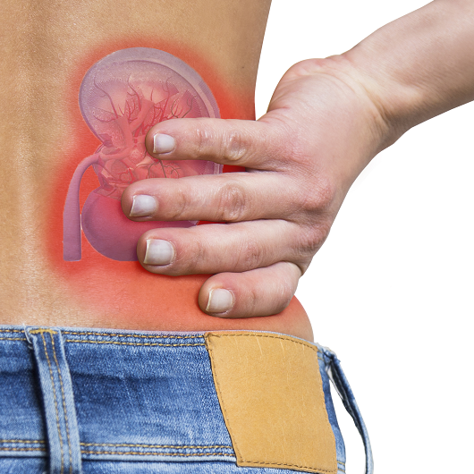
Can a Kidney Stone Go Away on its Own?
Kidney stones may pass naturally. Learn about treatment options from JFK University Medical Center urologists. Call 800-822-8905 for an appointment.

Mom of 3 Is Active and Carefree After Incontinence Treatment
After three vaginal births, Imani Waldman’s life was burdened with troublesome leakage. Then, the urogynecology team at Old Bridge Medical Center performed the life-changing Bulkamid procedure.

Should Women See a Urologist?
Women's urinary problems? JFK Medical Center urologist Dr. Lasser offers expert care for UTIs, incontinence, and more. Learn more and schedule an appointment today.