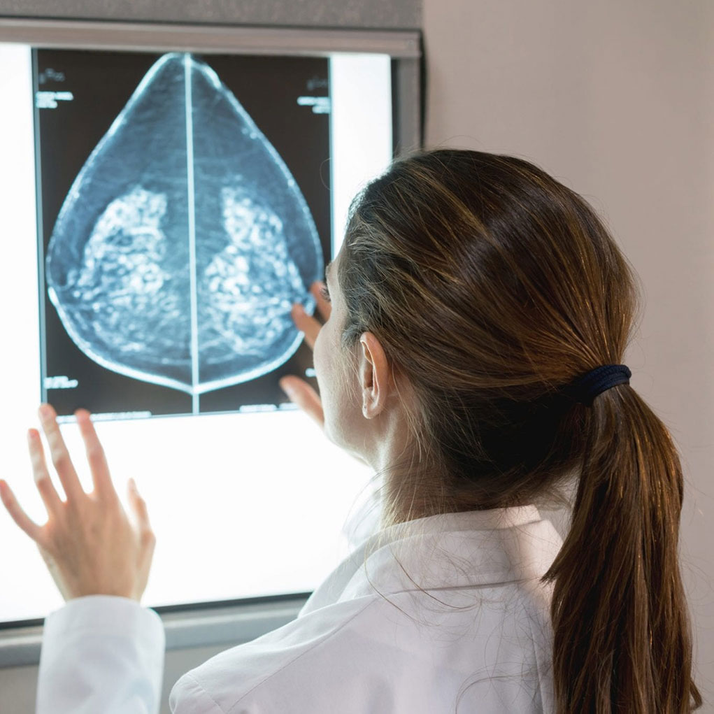
As a nurse practitioner, Madeline Rodriguez Hirsch understands the importance of proactive health care, which is why she was glad that the screening mammography order written by her gynecologist, Mark Brescia, M.D., included a supplemental screening through a breast ultrasound, in addition to the traditional mammogram.
“I had a history of prior benign breast biopsies, and there was always a concern that something else could be found,” Madeline says.
Adds Dr. Brescia, “Alone, breast ultrasound isn’t the best breast cancer screening method. But when used as an additional screening method in patients with dense breasts, ultrasound can find additional cancers that aren’t detectable on screening mammograms alone.”
Madeline’s anxiety was alleviated when she arrived at Palisades Medical Center. “I was greeted politely, and the radiology technologist performed the mammogram with great skill, knowledge and empathy,” she added. Not only was the radiology technologist gentle during the process, but she listened carefully to Madeline’s concerns about her previous need for follow-up.
Her experience with the ultrasound was equally as reassuring. “I was there late in the day because I came after work, but the ultrasound tech stayed to perform the ultrasound,” says Madeline. “Throughout the breast ultrasound, she explained exactly what she was doing and why, which made me less nervous.”
During Madeline’s follow-up mammogram, suspicious calcifications were identified and a stereotactic guided core biopsy was recommended. A stereotactic biopsy is an outpatient procedure that is simple and fast, and can eliminate the need for a surgical biopsy. It requires only local anesthesia and uses a small needle to very precisely extract breast tissue for further testing. Madeline promptly made an appointment for the biopsy.
The stereotactic breast biopsy was performed by Regina W. Chu, M.D., a diagnostic radiologist specializing in breast imaging at Palisades. “Dr. Chu performed the biopsy with care, explaining everything as she did it, allowing me to ask questions during the procedure,” Madeline says.
The biopsy brought fortunate results: The findings were benign.
Screening Mammograms vs. Diagnostic Mammograms
Current guidelines for average-risk patients recommend annual screening mammography beginning at age 40. In addition to annual screening mammography, some patients with dense breasts will need supplemental ultrasound screening. For high-risk patients, screening may need to begin earlier with additional screening modalities such as ultrasound or MRI. Approximately 10 percent of patients will be asked to return for additional imaging.
“There is a difference between screening and diagnostic mammograms,” Dr. Chu says. A screening mammogram is performed to detect breast cancer in an asymptomatic patient. When an abnormality is detected on a screening mammogram or if a patient presents with a clinical finding such as a palpable lump, focal pain or nipple discharge, a diagnostic mammogram is performed. A diagnostic mammogram can also be performed as a follow-up examination.
The diagnostic mammogram is monitored by a radiologist and will include additional views with specific attention to the area of the abnormality or to the area of clinical concern. For diagnostic workups, some facilities such as Palisades will accommodate same-day ultrasounds and, if there are findings on the ultrasound, same-day ultrasound biopsies for patient convenience.
The material provided through HealthU is intended to be used as general information only and should not replace the advice of your physician. Always consult your physician for individual care.


